 Graduate Microanatomy, 1998
Graduate Microanatomy, 1998
|
Respiratory system study guide Lab Exercises:
Date page was last edited 06/08/04 |
Laboratory Exercises:
Respiratory system Look at slide 48. The differential staining of this section shows the amount and distribution of elastic and collagenous fibers, smooth muscle, basement membranes, epithelium, cartilage, blood vessels, pleura, alveolar macrophages and capillary walls. The alveoli are collapsed in many regions. Look at the Pleural covering which shows a red-colored mesothelial lining. This is supported by connective tissue containing abundant collagen fibers stained green.
Find the smooth muscle in the wall of the bronchus. The fibers are arranged circularly and stained pink. Prominent features of the bronchial wall are the cartilaginous bars and plates. You can also see Bronchial vessels and nerve fiber bundles. Bronchi are accompanied by branches of the Pulmonary arteries. Note that the arteries and veins contain a considerable amount of elastic fibers.
Look at slides 46 and 47 for more structures in the lung. The lung section in slide 46 is partially covered by Pleura. Slide 46 shows air passages smaller than bronchi. These include Terminal Bronchioles, Respiratory Bronchioles, Alveolar Ducts, Alveolar Sacs and Alveoli. Find each of these structures on your slide 46, using the following photos as a guide.
Most broncioles show a pronounced smooth muscle coat. In this slide, the smooth muscle is red, embedded in green connective tissue. The bronchioles are also accompanied by pulmonary arteries, seen in the photograph to the left. Note that the bronchiole is easily distinguished from the thin walled alveoli. As you study these sections, you can appreciate the extent of the capillary bed in the lung. All blood vessels are filled with red blood cells, which delineate the capillary spaces. Occasional blood cells have spilled out....this is a fixation or postmortem artifact.
1) What is the function of the Clara cell?
Find both terminal bronchioles and respiratory bronchioles in this section. Recall that respiratory bronchioles have outpocketings of alveoli in the wall (hence their name). Terminal bronchioles, such as the one shown in the photograph to the left, end and connect with the walls of alveoli. In this photograph, the alveoli are arranged in a long duct and then they expand to form an alveolar sac. Find the same structures on your slide.
Another type of alveolar cell is the Type II cell. These are characterized by a reddish, foamy or vacuolated cytoplasm. Usually they are located at the angular junctions of alveolar walls.
Look at Slide 47. In this slide you should be able to see small Bronchi. You can also see bronchioles which are distinguished by the absence of cartilage plates. Identify terminal and respiratory bronchioles, alveolar ducts and alveolar sacs. Throughout the section, you may see particle-laden alveolar macrophages (shown in the following photograph).
2. What is the function of the Type II Alveolar cells?
3. Why do the ciliated cells extend further down the bronchiolar tree than the Goblet cells?
4. Define or draw the cellular and connective tissue layers which separate blood from the air spaces.
Return to top of page | Course Design | Learning Aids | Learning modalities | |
 Bronchus Find a bronchus in slide
48. Study its epithelium which is ciliated, respiratory epithelium with Goblet
cells. The epithelium rests on a purple-stained Basement Membrane.
Bronchus Find a bronchus in slide
48. Study its epithelium which is ciliated, respiratory epithelium with Goblet
cells. The epithelium rests on a purple-stained Basement Membrane.  Look at the epithelium under a higher power. Note the thick purple
stained basement membrane under which is a layer of black-stained, mostly longitudinally
arranged elastic fibers.
Look at the epithelium under a higher power. Note the thick purple
stained basement membrane under which is a layer of black-stained, mostly longitudinally
arranged elastic fibers.  The larger bronchioles are lined with an epithelium
that is pseudostratified columnar with or without cilia. However, the Goblet cells are no
longer present. Also, there are no more cartilage plates. As the bronchioles become
smaller, the epithelium becomes cuboidal.
The larger bronchioles are lined with an epithelium
that is pseudostratified columnar with or without cilia. However, the Goblet cells are no
longer present. Also, there are no more cartilage plates. As the bronchioles become
smaller, the epithelium becomes cuboidal.  This photograph shows a higher magnification of the
wall of a bronchiole. You can see the capillaries as well as the smooth muscle in
the wall. This is a small bronchiole because its epithelium is more cuboidal.
This photograph shows a higher magnification of the
wall of a bronchiole. You can see the capillaries as well as the smooth muscle in
the wall. This is a small bronchiole because its epithelium is more cuboidal. 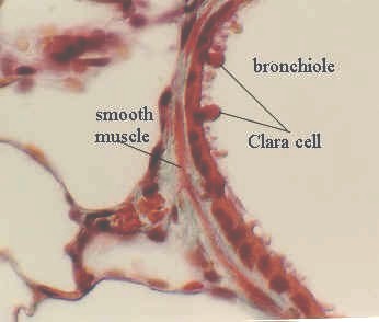 A higher magnification of a bronchiole wall shows the
ciliated cuboidal epithelial cells. There are also some non-ciliated rounded
secretory cells called Clara Cells. These are distinguished by their smooth rounded
apical "domes" which project into the bronchiolar lumen. Note again the smooth
muscle in the wall of the bronchiole.
A higher magnification of a bronchiole wall shows the
ciliated cuboidal epithelial cells. There are also some non-ciliated rounded
secretory cells called Clara Cells. These are distinguished by their smooth rounded
apical "domes" which project into the bronchiolar lumen. Note again the smooth
muscle in the wall of the bronchiole.
 A higher magnification of a terminal bronchiole shows
the transition between the bronchiolar wall and the alveolar walls.
A higher magnification of a terminal bronchiole shows
the transition between the bronchiolar wall and the alveolar walls. Look at the wall of alveoli. The Type I
alveolar cells are the extremely thin (0.2 microns) squamous cells.
Sometimes, only their nuclei are visible with the light microscope.
Look at the wall of alveoli. The Type I
alveolar cells are the extremely thin (0.2 microns) squamous cells.
Sometimes, only their nuclei are visible with the light microscope.  Two more Type II cells are shown in the photograph to
the left. The photograph beneath it shows more examples of the alveolar wall.
Two more Type II cells are shown in the photograph to
the left. The photograph beneath it shows more examples of the alveolar wall.
