

Seminiferous Tubules
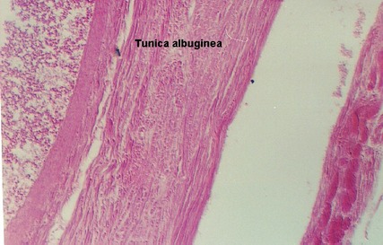
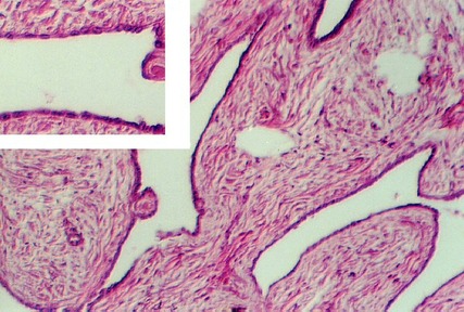
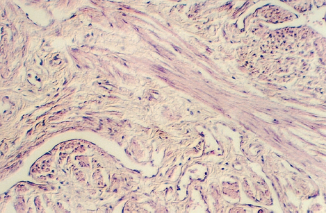
Study a section through the Testis and Epididymus. With the lowest power, identify the CREMASTERIC FASCIA outside the testis, containing skeletal muscle fibers. It is surrounded by connective tissue. Outside the muscle layer is the OUTER SPERMATIC FASCIA; inside the muscle fiber layer is the INNER SPERMATIC FASCIA. The next layer is an empty space. This is the cavity of the tunica vaginalis, derived from the embryonic processus vaginalis. Then, one encounters a layer of dense connective tissue called the TUNICA ALBUGINEA. It contains numerous large blood vessels. Inside the tunica albuginea one can find the numerous SEMINIFEROUS TUBULES. The following Figure shows, from left to right, the Tunica albuginea (with a blood vessel), the cavity of the tunica vaginalis, and some of the spermatic fascia.
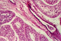
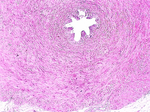
Look at the middle of the section of the Testis where there is connective tissue radiating outward from the MEDIASTINUM TESTIS. These connective tissues septa divide the testis into wedge-shaped lobulues. The Mediastinum is dense connective tissue in the middle of the gland. It is distinguished by channels lined by low cuboidal epithelium called RETE TESTIS. The following sections show the Rete testes.
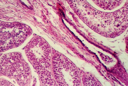
Seminiferous tubules contain the SPERMATAZOA in various stages of development along with the SERTOLI CELLS. Each tubule profile may show particular developmental stages in a high concentration, because development tends to occur in waves down the tubules. After the sperm are produced they travel to the TUBULI RECTI (straight tubules) which are found in the septa radiating from the mediastinum (see below). The tubuli recti connect the seminiferous tubules with the rete channels and tubules. The following photograph shows seminiferous tubules surrounding a connective tissue septum containing a tubulus rectus.
Sertoli Cells
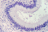
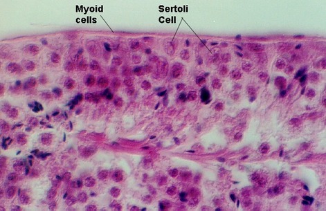
SERTOLI CELLS are distinguished by their location (near the border) and the shape of the nucleus. In the above photograph, note the outer layer of cells which are flattened and contractile (called MYOID CELLS). The Sertoli cell is identified by its triangular nucleus (kite-shaped). What is the function of the Sertoli cell?
Spermatogonia
SPERMATOGONIA are also close to the basal lamina. They are large round cells with a pale-staining round or oval nucleus that is distinctly different from the nucleus of Sertoli cells. Sometimes you can see them in mitotic division. Can you find a Sertoli cell in the above photograph?
Spermatocytes
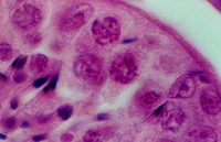
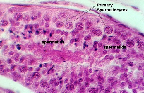
In the next rows, further away from the periphery, are either PRIMARY SPERMATOCYTES or SPERMATIDS. The Primary spermatocytes have a large floccular nucleus. They can be found in the following photograph.
Spermatids
Similar, but smaller secondary spermatocytes might be seen, however, because they complete meiosis II rapidly, you are more likely to see the smaller early spermatids. The early spermatid is small and round whereas the LATE SPERMATID is more conical. It may have a flagellum and a dense nucleus. The above photograph shows both primary spermatocytes and spermatids. Spermatids are shown also in the following photographs.
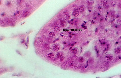
In the following photo, you should be able to find Sertoli cells, Primary spermatocytes, and spermatids. Identify an example of each.

Leydig Cells
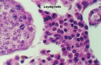
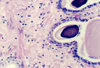
The connective tissue in-between the tubules contains clusters of INTERSTITIAL CELLS (LEYDIG CELLS). They will be difficult to find in slide 72. You can, however, see them in slide 4. This slide does not have well preserved seminiferous tubules, however the Leydig cells are prominent. The following two sections illustrate them. They are round, acidophilic (pink) and may contain some vacuoles.

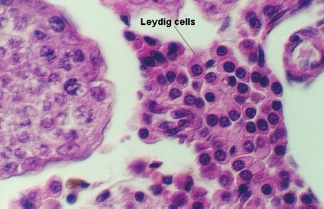
What is the function of the Leydig cell? What hormone does it secrete? What hormone stimulates Leydig cells and where is that hormone secreted?
The following photograph shows a section through a fetal testis. From your studies above, you should be able to distinguish Leydig cells and Seminiferous tubules.

In the seminiferous tubules, what is the most frequent cell type found? What other cell type might be found and why?
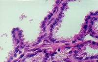
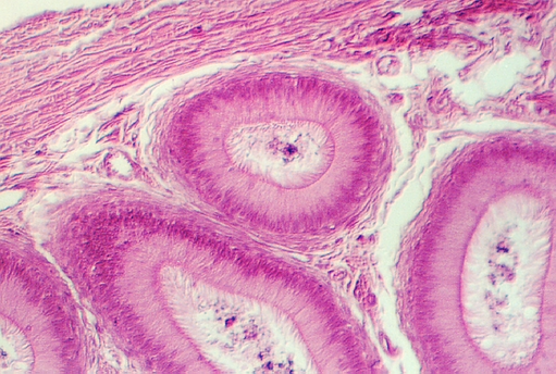
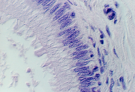
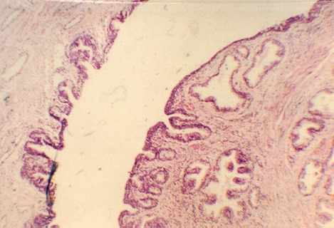
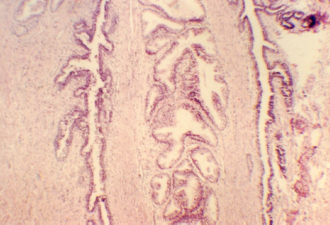
Look at slides 5 and 72 for sections through the epididymus. The first set of tubules you need to find, however are the EFFERENT DUCTULES. These are lined with a low pseudostratified epithelium that is very ragged. Sometimes the tubules look "festooned". The efferent ductules receive sperm from the Rete testes channels. Then they merge to form the single duct of the EPIDIDYMUS. Consult an overview showing the organization of the male reproductive system. Efferent ductules are shown in the following photos.
This low magnification shows the ragged, festooned appearance of the Efferent ducts.
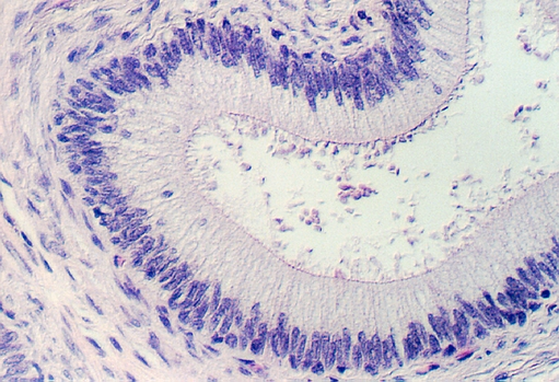
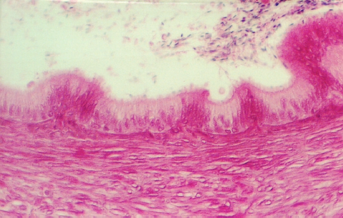
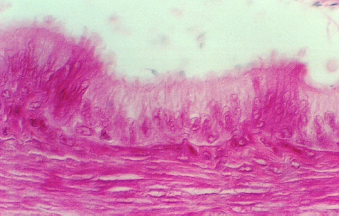
The low pseudostratified epithelium is shown in the following photos.

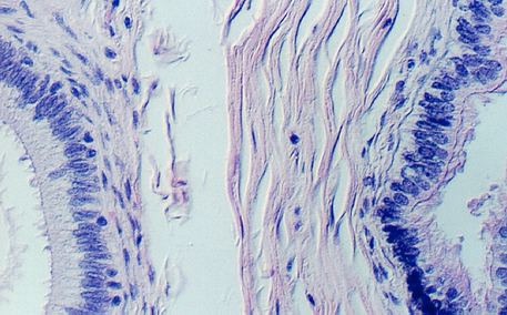
As the Efferent ductules merge, the epithelium gradually changes to simple columnar with tufts of STEREOCILIA. The following photograph shows a higher efferent ductule on the right and an epididymal duct on the left.
Epididymus
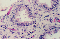
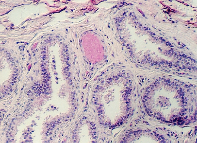
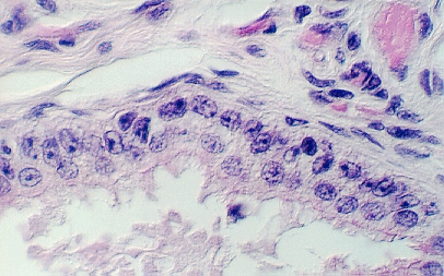
The main portion of the duct of the epididymus is characterized by high columnar epithelium distinguished by sterocilia which reach into the lumen of the duct. The nuclei of these cells, called PRINCIPAL CELLS, are elongated and located at the base. Another population of cells has a rounder nucleus and can be found mainly at the base (called BASAL CELLS). These are considered 'reserve cells' for the principal cells. You can find epididymus ducts on both slides 5 and 72. The illustrations below should help you.
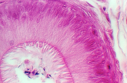
Identify principal and basal cells in one of these photos. Identify the stereocilia in the following photograph. Are there microtubules in stereocilia? What is their function?
What is the major function of the epididymus?
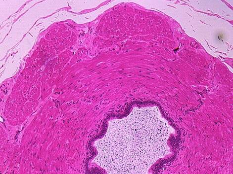
After the sperm leave the epididymus, they travel down the vas deferens (ductus deferens). These highly muscular tubes are sensitive to oxytocin and very important during ejaculation. The DUCTUS DEFERENS or Vas deferens is seen in slide 72 and also in slide 73. Some of the sections may have the epithelium destroyed. Try to find one that has intact epithelium. Below are photos from slide 72. The lumen is lined by a pseudostratified columnar epithelium that may also have sterocilia. Sometimes the epithelium has complex infoldings. The stereocilia gradually disappear towards the distal regions of the duct.
The smooth muscle fibers of the wall are arranged principally in an inner longitudinal, middle circular, and an outer longitudinal direction. The photos below (not from your class slide set), illustrate the three layers.
The Vas deferens penetrates the Prostate gland and becomes the ejaculatory duct. There are two ejaculatory ducts, one from each side.
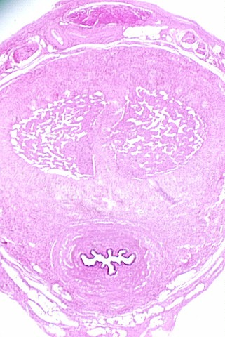
In order to see the prostatic urethra, be sure and find a slide that shows the entire gland. Hold that slide up to the light and observe the U-shaped lumen of the urethra in the center of the prostate. The hill like tissue mass forming the concave curve of the urethra is called the COLLICULUS SEMINALIS. In that tissue mass, look for two EJACULATORY DUCTS. These represent the junction points for the vas deferens from each epididymus. They are distinguished by their dense epithelium which looks clearly different from that of the gland. The photograph below shows the curving urethra. The ejaculatory ducts will empty their content of sperm into the prostatic urethra.
The following photograph shows the two ejaculatory ducts in the colliculus seminalis. They lie to either side of glandular tissue and show a distinctive dense epithelium.
Higher magnifications of the ejaculatory ducts are seen below. Note that the colliculus seminalis tissue has a great deal of smooth muscle. This will no doubt contract to help empty the sperm into the prostatic urethra.
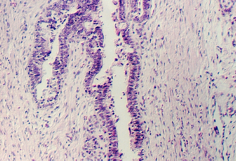
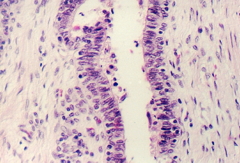
Diagram or summarize the pathway that sperm take from the Seminiferous tubules to the urethra. Trace the urethra to the penis
PENIS: Penile urethra
First, hold this slide to the light and look for the urethra, an opening towards one end. Then, use low power to identify a number of landmarks. First, the epidermis should be obvious with pigmented basal layers containing numerous melanin granules. You may also see occasional sweat and sebaceous glands. Another striking feature is the abundant smooth muscle and nerves, just under the skin.
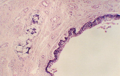
With a low magnification, you can see the dense connective tissue layer that surrounds each of the CORPORA CAVERNOSA and is continuous with the septum penis between the two corpora cavernosa. The photo shows a low magnification view of a section through a fetal penis. The two corpora cavernosa are evident subdivided by the connective tissue septum. You can also see the penile urethra at the bottom of the section. It is in the corpus spongiosum.
Find the urethra on your slide. It is surrounded by erectile tissue called the CORPUS SPONGIOSUM. The epithelium may not be well preserved. Occasional urethral glands (GLANDS of LITTRE) can be seen. They are mucous glands. The following photograph shows a section through the penile urethra. There are glands of Littre in the connective tissue under the epithelium.
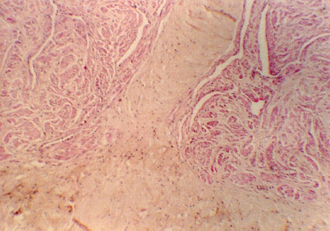
A higher magnification of the septum separating the two corpora cavernosa is shown in the following photo. The well vascularized erectile tissue is seen in the corpora as well as in the next photo (higher magnification.
http://microanatomy.net/Male_Reproductive/testes_and_ducts.htm
Gwen V. Childs, Ph.D., FAAA
Department of Neurobiology and Developmental Sciences
University of Arkansas for Medical Sciences
4301 W. Markham, Slot 510, Little Rock, AR 72205
For questions or concerns, send email to this address
