The liver slides in the class slide collection include sections stained different ways. You will find liver sections on slides 59, 60, 61, 26, and 2. Some characteristics are seen more easily on one or more of the sections. We will highlight those as we review the liver slides.
Slide 59 is an H&E stained section of the liver and slide 60 is a Trichrome stained section. Use both of these slides to identify most of the features in the liver. Note that the liver is covered by a connective tissue capsule called GLISSON's CAPSULE. The HEPATIC LOBULE is defined by a radiating arrangement of liver cell plates around a CENTRAL VEIN.
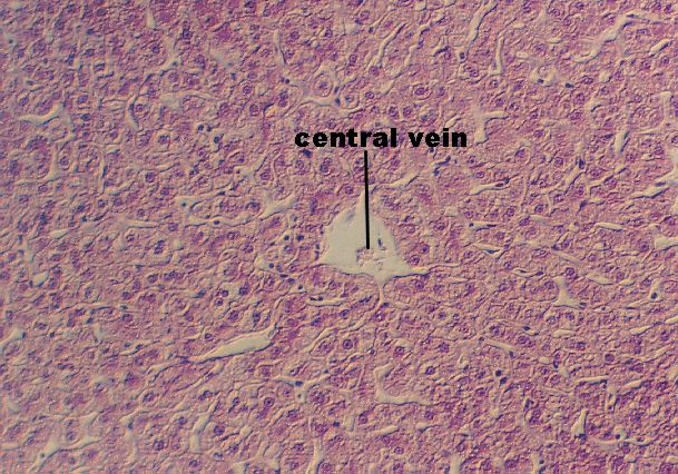
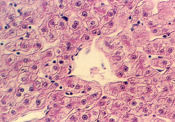
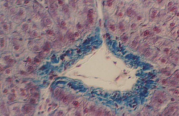
The hepatic parenchymal cells may be binucleate (can you find one in the above photo?). On either side of the rows of liver cells, are the sinusoids. Venous blood that will leave the liver flows towards the central vein (center of the Hepatic lobule.) Another view of the Central vein, from slide 60, is shown in the following photograph. You can see the connective tissue wall of the vein better because the trichrome stain is bright blue.
When you study the liver, consider how the liver cells receive arterial blood. Name the vessels and trace the blood to the Central Vein. They also receive blood from the intestine. Trace that supply as well and understand the significance of the dual sources. The sinusoids actually contain a mixture of blood from the two supplies.
Kupffer Cells and Sinusoids
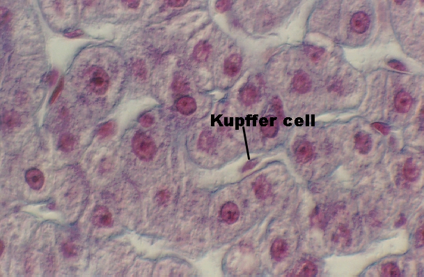
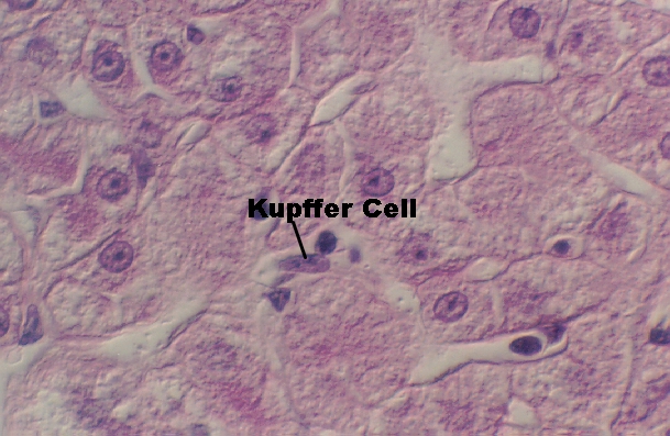
Stretching across the SINUSOIDS are phagocytic cells called KUPFFER CELLS which are distinguished by their processes. The following photographs show Kupffer cells from slides 60, 59, and 26.
As you study the structures, be sure and define the SPACE of DISSE?
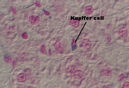 The Kupffer cell to the left was distinguished after
the animal was exposed to trypan blue. It took up the dye and its cytoplasm is blue.
The Kupffer cell to the left was distinguished after
the animal was exposed to trypan blue. It took up the dye and its cytoplasm is blue.
Bile Canaliculi and Bile Duct
Bile flows from the liver cells out through small channels between the cells called BILE CANALICULI to the BILE DUCTS located along the sides of the Hepatic lobule. These can be seen in thin plastic embedded sections of the liver in slide 2. They appear as a small round gap between adjacent hepatocytes. The SPACE of DISSE can also be seen, however it is artifactually enlarged.

The Portal Lobule
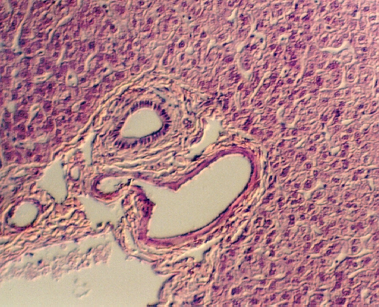
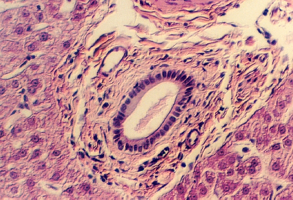
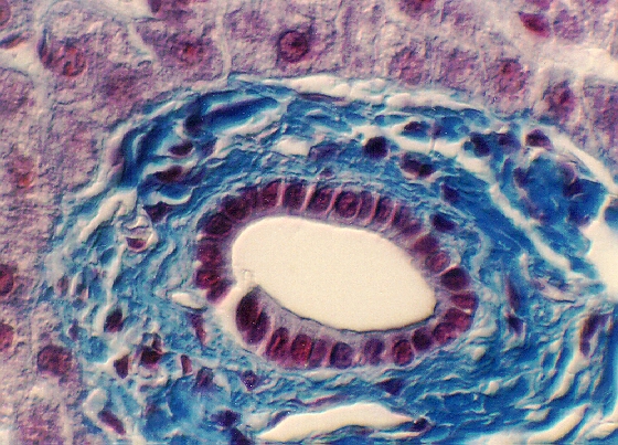
One can also define liver lobules functionally from the direction of bile flow. A PORTAL LOBULE is represented as that liver tissue with the BILE DUCT, HEPATIC ARTERY and PORTAL VEIN in the center. Collectively, these three structures are called the TRIAD. In the following photographs, you can see the Hepatic artery, Portal vein, and the Bile duct which is distinguished by its cuboidal epithelium. These are from slides 59 and 60. The branches of the hepatic arteries and portal veins are not easy to find in the triads in slide 60, because of fixation problems.
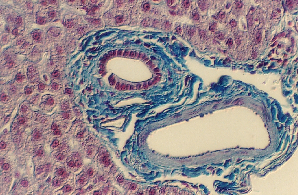
Look at slide 61. This was stained with a van Giesson Stain. The connective tissue is stained red, the cytoplasm yellow, and the cell nuclei brown. BILE DUCTS can be seen. HEPATIC Arteries can also be distinguished by the yellow brown smooth muscle in the arterial walls. You can also find CENTRAL VEINS.
Recall the source of the blood in the portal vein. Trace it from its origin. Why is it important for liver function?
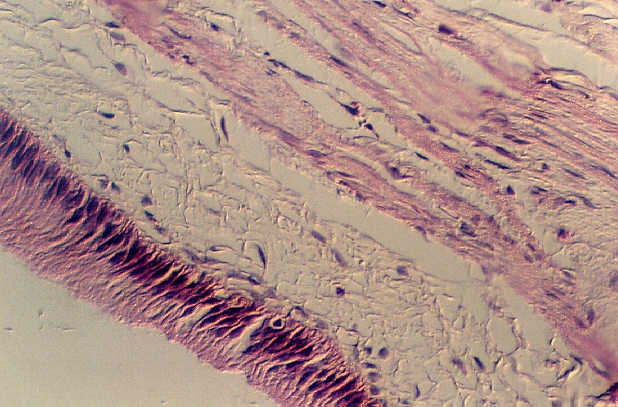
Examine slide 62. There are two sections, one with a distended gall bladder and flattened mucosa. The other shows an empty gall bladder. Note that the MUCOSA is stretched out in the distended gall bladder and thrown into many folds in the empty organ. The epithelium is simple columnar with a small brush border. There is no muscularis mucosa. Underneath the epithelium is the submucosa. The muscularis externa consists of irregularly interwoven strands of smooth muscle, collagenous and elastic fibers. The following figure shows the distended gall bladder.
Trace the flow of bile from the liver to the gall bladder. What is the function of the gall bladder?
Slide 58 shows the Pancreas. It is surrounded by a thin connective tissue CAPSULE from which SEPT extend into the interior, thereby subdividing the parenchyma into smaller units called LOBES and LOBULES.
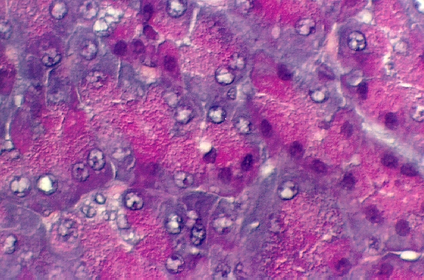
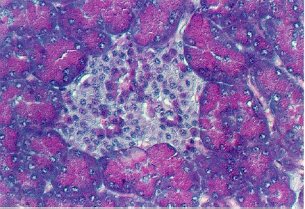 The pancreatic tissue is composed of exocrine ACINI.
The apical portions of their cells contain an abundance of zymogen granules which
are stained bright red. The base is more basophilic, because of the rough endoplasmic
reticulum.
The pancreatic tissue is composed of exocrine ACINI.
The apical portions of their cells contain an abundance of zymogen granules which
are stained bright red. The base is more basophilic, because of the rough endoplasmic
reticulum.
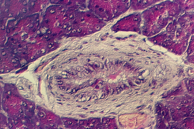
Large EXOCRINE DUCTS are seen in the connective tissue. The largest EXCRETORY DUCTS contain a conspicuous amount of SMOOTH MUSCLE. The epithelium lining the ducts is SIMPLE to PSEUDOSTRATIFIED COLUMNAR.

Where do the digestive enzymes go when they are secreted? What prevents them from digesting the wall of the pancreatic ducts?
Return to top of page
Last updated: 02/21/11
© copyright 1998 Gwen V. Childs, Ph.D.
URL Address: http://www.microanatomy.net/digestive/Liver_and_pancreas.htm
Gwen V. Childs, Ph.D., WebMistress
childsgwenv@uams.edu