

SPECIALIZED CELLS IN CONNECTIVE TISSUE
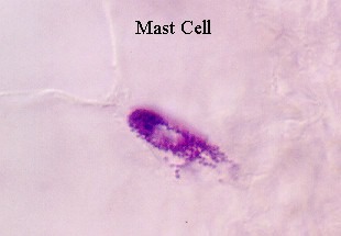
Mast cells on a smear of connective tissue stained to help you identify the mast cells. They are seen by their granules, containing histamine which is released after an allergic reaction. How is histamine triggered and what does it do?
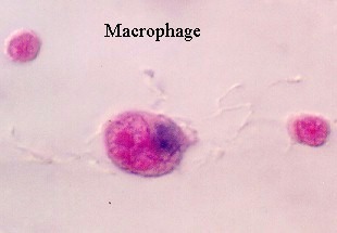
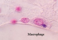
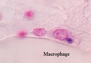
These photographs show a connective tissue preparation designed to visualize the macrophages.
These cells have been exposed to trypan blue, while living and the macrophages have brought in the dye by phagocytosis, allowing them to be identified. Nuclei are stained light pink, in this preparation.
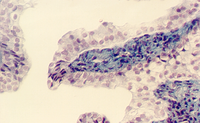
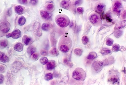
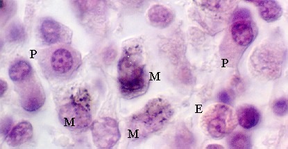
The following photographs show two important cells in loose connective tissue. The inner regions of the lymph node called the medulla, is filled with connective tissue that contains numerous macrophages (M), eosinophils (E) and plasma cells (P). The macrophages can be identified by their content of large granules. The eosinophils have pale or bright red granules and a lobed nucleus. Plasma cells can be identified by their ovoid shape, nucleus that is off-center, and nuclear chromatin pattern which looks like a "cartwheel". Sometimes there is a pale area like a half moon near the nucleus. This is the Golgi complex. What is the function of a plasma cell?
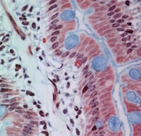
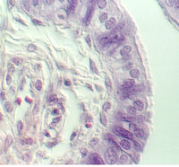
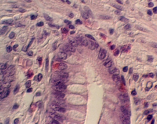
One of the types of white blood cells, called an eosinophil is found in loose connective tissue and may increase in certain types of inflammatory responses. What types of inflammatory responses would you expect to bring more eosinophils? These cells are distinguished by their bright red granules and their multilobed nuclei. You can see them in intestine or lymph node slides. Note that in this slide there are numerous eosinophils in the connective tissues under the epithelium. However, two more cells with red granules can be found in the epithelium (can you find them?). These are not eosinophils. They are specialized endocrine cells that secrete hormones and growth factors important to the GI tract (called "enteroendocrine cells".)
URL Address: http://microanatomy.net/connective_tissue/specialized_cells.htm
Gwen V. Childs, Ph.D., FAAA
Department of Neurobiology and Developmental Sciences
University of Arkansas for Medical Sciences
4301 W. Markham, Slot 510
Little Rock, AR 72205
